> Experimental Work > Clinical Research

Clinical Research

脳動脈瘤破裂機構の解明
脳動脈瘤が破裂するメカニズムは未だに不明です。これを解明すべく、臨床研究と基礎実験の両面から研究しています。手術で摘出した動脈瘤標本に対して、走査電子顕微鏡や免疫染色で動脈瘤壁を観察するとともに、術前3D画像から流体解析を行い、力学的要素と病理学的要素を関連づけて解析しています。併せて、旧来型ラット脳動脈瘤モデルを改変して容易に破裂する大型動脈瘤モデルを作り、薬剤投与実験を行っています。この研究が実れば、手術に頼らずに内科的な方法でクモ膜下出血を防止することができるようになるかもしれません。 
image21


Investigating brain networks in injured brain
Brain injury from neurotrauma, cerebral vascular diseases as well as tumor involves multiple brain networks.  Recent development of neuroimaging postprocessing technique as well as statistical analysis enabled noninvasive measurements of brain morphology as well as functions in the brain areas.  Nevertheless, investigating brain networks is challenging in the brain with injury, and it will only be possible none other than neurosurgeons.  Our goal is to provide optimal strategy for the surgical intervention in patients with Moyamoya disease.  Since a certain number of patients suffer from cognitive impairments, the timing of revascularization as well as medical intervention should be optimized. Research of brain structure and function in patients with moyamoya disease will provide a novel insight into vascular cognitive impairments as well as makers to track convert ischemic injury.
image22


EZO (effective zone for mobile stroke team) trial 
A treatment for cerebral infarction has been progressed by an advancement of endovascular treatment in recent years, but because of the fewer number of doctors available for a new treatment, the treatment may not be appropriately provided in some regions. Thus, in cooperation with the related facilities, Hokkaido University Hospital has established a system where medical specialists can provide treatment supports by traveling. If this treatment support system is scientifically proved as effective, it will give a greater chance to provide appropriate medical services to more people <See Figure>. Since there is a concern that regional gaps in terms of medical quality would increasingly be further widening in future, it is expected that the system could be one of the methods to solve such issues.
image23


Measurement of blood viscosity by an electromagnetic spinning sphere (EMS) viscometer in ischemic stroke patients
Although increased blood viscosity (BV) has been identified in the in the acute phase of ischemic stroke, actual measurements of BV have not been performed in clinical practice recently. We attempted to reconfirm the meaning of measurement of BV in acute ischemic stroke patients classified into ischemic stroke subtypes by using an electromagnetic spinning sphere (EMS) viscometer (Cooperation study with Prof. Sakai who developed the EMS viscometer in Institute of Industrial Science, the University of Tokyo).
The results of the measurement revealed that BV did not equally increase in all stroke subtypes, and it significantly increased in patients with small artery occlusion (SAO) at the date of admission, suggesting the involvement of dehydration. From now on, we would like to analyze BV values more in detail by investigation of shear-rate dependency and comparison between whole blood viscosity and plasma viscosity.
image24


Usefulness of 11C-methionine PET for brain tumors
Positron emission tomography (PET) is now one of the essential examinations in the treatment of cancer, and especially, 11C-methioine (MET) PET have a great potential for the diagnosis of brain tumors. In present, we have two open-labeled clinical research of MET PET for brain tumor with the Department of Nuclear Medicine under the “advanced medical care” system. One clinical research is whether MET PET can distinguish tumor recurrence from radiation necrosis. Radiation therapy is critical treatment for malignant brain tumors, but some patients would suffer from radiation necrosis. It is difficult to distinguish between real recurrence and radiation necrosis by MRI or FDG PET, but MET PET have a potential to distinguish real recurrence from radiation necrosis. The other clinical research is whether MET PET can detect the tumor infiltrative area in newly-diagnosed glioma. Both clinical research have received a lot of attention recently.
image25


個別化医療に向けた脳腫瘍バイオバンクの確立
がん治療において、従来のような疾患別の治療法ではなく、腫瘍特有の遺伝子異常(体細胞変異といいます)に基づいた個別化医療が実現しつつあります。近年の大規模ゲノム解析により、脳腫瘍においても特有の遺伝子異常が徐々に判明しています。近い将来、脳腫瘍治療においても個別化医療が導入されることを想定し、その基盤作りのためのバイオバンク確立を行っています(病理・遺伝子診断科との共同研究)。また、このバイオバンクで得られた情報を基にした更なる臨床研究も計画中です。


Activities as a pediatric cancer center
apanese Ministry of Health, Labor and Welfare specified Hokkaido University Hospital as one of the pediatric cancer centers. We work on many clinical researches in collaboration with Japan Children’s Cancer Group, for example, Genome Wide Association Study concerning germ cell tumors of CNS and Molecular diagnosis research of pediatric brain tumor.

Proton beam therapy for patients of pediatric brain tumor
We have the newly opened facility for proton beam therapy. Proton beams display a feature to release the main dose of energy close to the tumors, and the surrounding normal tissue is not affected. It offers numerous benefits to patients of pediatric cancer. We have a weekly pediatric cancer board with department radiation oncology and pediatrics.


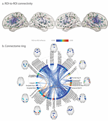
Consideration of high order brain function after deep brain stimulation for Parkinson disease
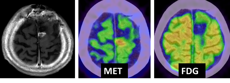
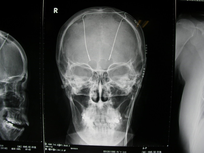
Deep brain stimulation, following DBS, is performed for involuntary movements, such as Parkinson disease, idiopathic tremor, so on (see figure). Moreover, it is well-known that some patients show mental disorder or cognitive disorders, such as depression, apathy, so on. However, it is controversial as for the mechanism. We will perform a prospective study as for high order brain function after DBS using various clinical psychological examination and functional imaging (Independent clinical research; 014-0032).
image26